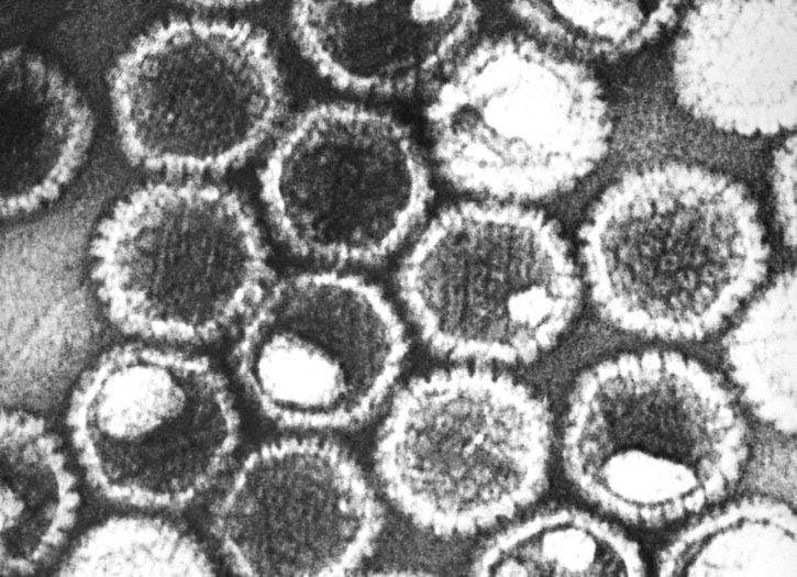
Researchers from the University of Glasgow have used Nobel prize-winning cryo-electron microscopy (cryo-EM) to reveal the molecular structure of the common herpes virus in high definition.
The capsid, which stores the virus’ DNA, has been difficult for scientist to analyse because of its microscopic size; it measures 1/10000th of a millimetre in diameter.

Discover B2B Marketing That Performs
Combine business intelligence and editorial excellence to reach engaged professionals across 36 leading media platforms.
Cryo-EM has enabled researchers to visualise and understand the structure and mechanism of a motor-like molecule, which they named the portal. It is used to pump the herpes virus’ DNA into the capsid for storage and to eject the DNA from the capsid when infecting the host’s cells. The technique also illustrated how the herpes virus packs DNA inside the capsid.
Dr David Bhella, lead author of the study from the MRC-University of Glasgow Centre for Virus Research, said: “Cryo-electron microscopy, combined with new computational image processing methods allowed us to reveal the detailed structure of the unique machinery by which the virus packs DNA into the capsid. The DNA is packed very tightly, reaching a pressure similar to that inside a bottle of Champagne.
“We hope that this study will eventually lead to the development of new medicines to treat acute herpesvirus infections, through the design of drugs that will block the action of the portal motor.”
Cryo-EM was developed by the Medical Research Council (MRC) Laboratory of Molecular Biology. It works on a frozen sample, which is studied by a beam of electrons. These electrons interact with the molecules and map the structure, movement and functions of biomolecules. The resulting image is more detailed and clear than those produced by alternative microscopy techniques.

US Tariffs are shifting - will you react or anticipate?
Don’t let policy changes catch you off guard. Stay proactive with real-time data and expert analysis.
By GlobalDataMRC, which funded this University of Glasgow study, head of infections and immunity Dr Jonathan Pearce said: “Dr Bhella and his team have now used this technique to elucidate the structure of the herpes virus, revealing a ‘molecular machine’ that is involved in virus replication. The findings provide scientists with a better understanding of the virus and its anatomy, and, in turn, an insight into potential new therapeutic targets.
“These elegant experiments exemplify the potential of the Scottish Centre for Macromolecular Imaging due to be launched later this year at the CVR, led by Dr Bhella, which will support vital research into diseases posing the greatest threat to human health.”
Members of the herpes virus family can cause both cold sores and chicken pox, as well as cancers and severe illness in unborn children. The University of Glasgow research team hope this breakthrough will aid the development of drugs to treat the disorder.


