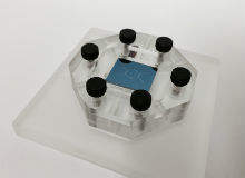

A breakthrough lab-on-a-chip technology developed by IBM Research scientists could enable doctors to detect diseases like cancer before symptoms even appear.

Discover B2B Marketing That Performs
Combine business intelligence and editorial excellence to reach engaged professionals across 36 leading media platforms.
The system, described in the journal Nature Nanotechnology, can separate biological particles down to 20 nanometers in diameter, a scale that gives access to important particles like DNA, viruses and exosomes. Once separated, these particles could then potentially be analysed by doctors to reveal signs of disease earlier than ever before and when outcomes for treatment are most positive.
After originally focusing on exosomes, which can be captured non-invasively by ‘liquid biopsies’ and are increasingly being viewed as useful biomarkers for malignant tumours, IBM’s next step is to confirm – with the help of a team from the Icahn School of Medicine at Mount Sinai in New York, US – that their device is able to pick up exosomes with cancer-specific biomarkers.
“By targeting exosomes, we anticipate our lab-on-a-chip technology can give physicians a view into the origin of a cancer or if a cancer has metastasized before physical symptoms appear in the patient,” explains Joshua Smith, an IBM researcher in the Nanobiotechnology Group and one of the leaders of the project.
Elly Earls meets Smith to find out why their new system, which can separate particles at a scale 50 times smaller than ever achieved before, has applications far beyond cancer, and how soon it might be available to patients.

US Tariffs are shifting - will you react or anticipate?
Don’t let policy changes catch you off guard. Stay proactive with real-time data and expert analysis.
By GlobalDataElly Earls: Please describe the technique you’ve developed at IBM to separate bioparticles down to 20 nanometers in diameter. How significant a move forward is this from current techniques?
Joshua Smith: Separation of nanoscale particles has been possible for years in various forms, albeit through much more cumbersome methods such as ultracentrifugation, gel electrophoresis, chromatography or filtration. The main disadvantage to these approaches rests in compromise: centrifugation and chromatography can be very precise but require expensive machinery and trained technicians, whereas gels and filter media are cheap and easy to use but are less precise and more difficult to recover samples from.
What we have done is to use IBM's vast knowledge and capabilities in silicon nanotechnology, applying it to healthcare, to dramatically scale a specific technology – deterministic lateral displacement (DLD) separation technology, where bioparticles are separated as they zig zag through pillar arrays on a chip – beyond what others have been able to achieve. We've taken the process down to the scale of a chip and begun to build a standardised platform where our separation capability can be coupled with detection, manipulation and other analytic capabilities that would prove helpful to a physician in a clinical setting.
EE: What have been the main challenges you’ve had to overcome along the way?
JS: Fabrication, nanofluidics and biochemistry were the main challenges the team had to overcome and all the authors were able to contribute to these areas of expertise. First off, it was a major challenge to make the pillar structure and gap sizes down to 25nm with high aspect ratios. This required innovations in process engineering.
Also, micro-fabricated silicon is not a natural environment for bioparticles, so a tremendous amount of work on varying the surface chemistry was required to achieve compatibility with exosomes.
Lastly, in operating at the nanoscale, new phenomenon emerge that are not present in microfluidic devices, and this required substantial testing and development of a physical model to both understand and control the separation process.
EE: The paper IBM published in Nature Nanotechnology showed that in principle your lab-on-a-chip technology can help with early disease detection by separating bioparticles, specifically exosomes, at the nanoscale. Why did you focus on exosomes?
JS: Exosomes are an emerging field for medical research as scientists have realised that they can reveal critical information about a disease, especially cancer. Traditionally, people considered exosomes as garbage cans of unwanted cellular material that were excreted by cells. In time, scientists have acknowledged that exosomes carry important genetic cargo such as DNA, RNA, surface proteins and other biomarkers that are transported between cells.
Exosomes can be found in abundance in bodily fluids such as blood, urine or saliva, and in principle, cancer cells are constantly shedding exosomes as they are very active. Therefore, by having a less invasive and cheap way of separating out exosomes for analysis, our lab-on-a-chip technology can give physicians a view into the origin of a cancer or if a cancer has metastasized before physical symptoms appear in the patient. Also, physicians may be able to use the technology as a point-of-care device to see how the patient is doing after treatment.
EE: What’s the aim of your collaboration with Mount Sinai?
JS: With Mount Sinai, we plan to confirm if our technology is able to pick up exosomes with cancer-specific biomarkers from patient liquid biopsies. Discovering the onset of early disease can be compared to finding a needle in a haystack, in that separating a specific type of bioparticle, such as a prostate exosome, from a complex biological mixture requires a methodical process to isolate that target.
This process has traditionally required expensive equipment, wet lab environment and trained technicians, making regular sample preparation and monitoring of disease progression impractical for most. Our technology has the potential to open the door to on-chip separation and purification at a scale that circumvents some of these previous requirements, enabling the possibility of cheap, load-and-go preparation – a precursor to any diagnostic.
EE: How long do you think it will be before your device, or something similar, can be used in a clinical setting? What challenges need to be overcome first?
JS: There are still many engineering challenges to overcome in order to improve the efficiency and volume needed to detect a sufficient number of exosomes that allow us to determine what cargo they carry from cancer cells or for other diseases. Eventually we want to be able to have a read-out of different biomarkers found among the exosomes that a physician could interpret in the context of the health of the patient.
We plan to immediately begin working with hospitals and researchers outside of IBM to help us develop and fine tune the technology. Our collaboration with physicians and researchers at Mount Sinai Hospital will provide the initial proving ground for the technology.
EE: What’s your ultimate goal?
JS: While the focus of the paper is centered entirely on exosomes, access to this size scale opens up the possibility of sorting other nanoscale bioparticles, such as viruses (Zika and flu) and nucleic acids.
The goal is to create an adaptable lab-on-a-chip platform through a broader integration strategy doing exactly what IBM has done very well historically, which is the development of advanced semiconductor platforms. We believe that harnessing IBM's expertise in this area together with our growing understanding of experimental biology will help us reach this goal.
EE: How affordable do you anticipate the device will be?
JS: We want to have a small and affordable diagnostic tool that can detect minute quantities of biomarker particles that tell physicians something about a person’s health. The ability for physicians to regularly screen an individual’s biological profile in an affordable and noninvasive manner has the potential to usher in a new era of preventative healthcare and further our understanding of the factors that contribute to disease progression.


